Abdominal Evaluation Is Performed in Which of the Following Sequences
Ing the following specific measures extracted from clin-ical abdominal images. Inspection consists of visual examination of the abdomen with note made of the shape of the abdomen skin abnormalities abdominal masses and the movement of the abdominal wall with respiration.
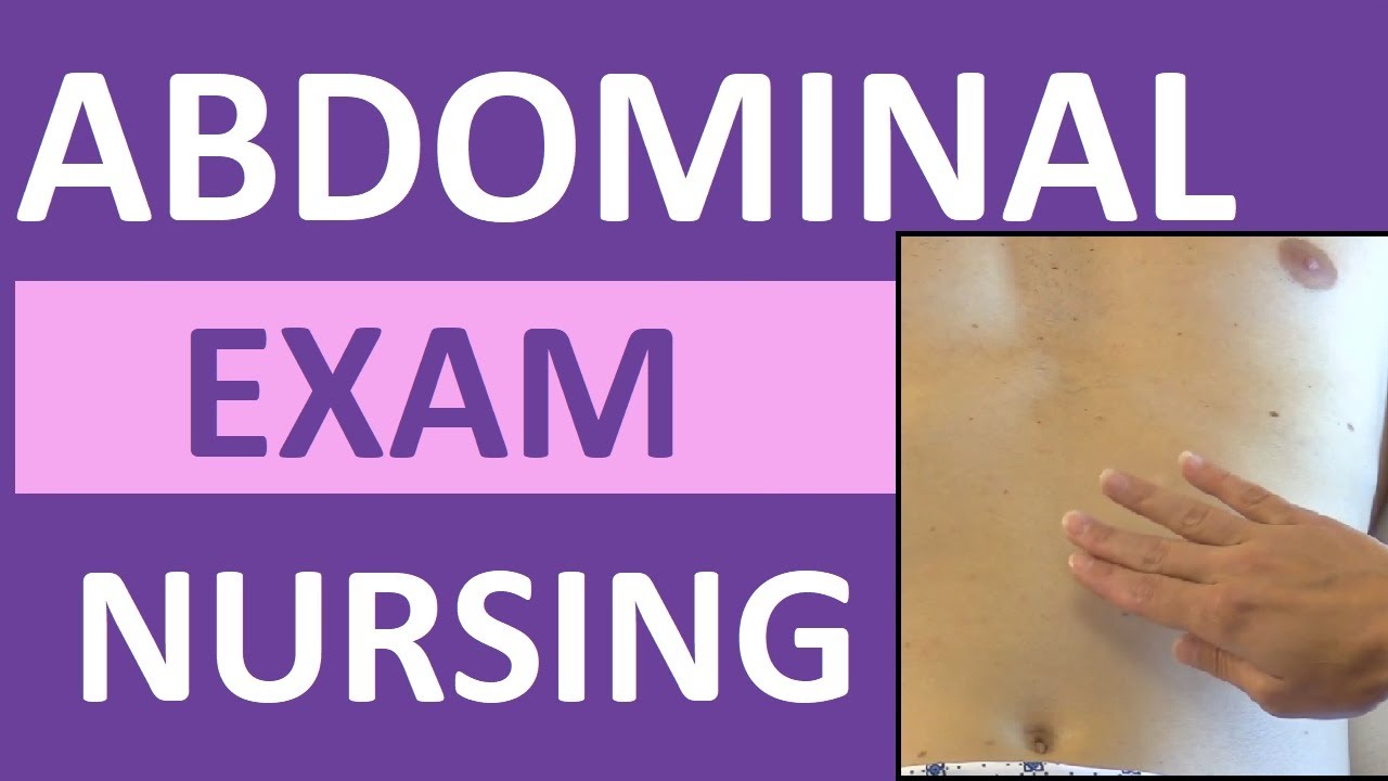
Abdominal Examination Exam Nursing Assessment Bowel Vascular Sounds Palpation Inspection Youtube
-Postoperative abscess -Acute inflammatory bowel disease exacerbation -Small bowel obstruction-Surveillance of low grade abdominal neoplasm Retrospective review to identify all patients.

. 4 These sequences can be. Magnetic Resonance Imaging in the Evaluation of Abdominal Aortic Aneurysm Martin R. The physical examination typically occurs after a thorough medical history is taken that is after the physician asks the patient the course of their symptoms.
The MRI protocol included fat-saturated T1-weighted spin-echo SE sequences pre- and post iv. It is currently one of the most powerful and versatile imaging procedure for the evaluation of abdominal masses. Which of the following procedures involves the evaluation of the most distal portions of the gastrointestinal tract.
When your intestines process food your abdomen may grumble or growl. Scratch marks pruritis is a feature of cholestatic liver disease Bruising due to impaired clotting factor production in liver failureS. Steady-state free precession sequences also termed balanced-FFE true-FISP and FIESTA are rapid have high intrinsic contrast resolution and have been shown to be accurate in evaluating renal artery stenosis 2 aneurysm sac contents Figure 102-1 3 thoracic aortic dissection and aneurysm.
The criteria for tumor manifestation was contrast enhancement of intra-abdominal soft tissue lesions or peritoneum. _____ sequences are performed to suppress CSF cerebro-spinal fluidand aid in. TSE sequences were performed with the HASTE sequence TR.
In the last 3 years 80 patients with abdominal aortic aneurysms identified with US or CT were examined with 05-T MRI and underwent surgical repair within 15 days. Analysis of the same MRI slices verified by tissue artefacts showed a statistically significant all P 00001. The following modifications are made.
The abdominal sounds you hear are most likely related to the movement of food liquids digestive juices and air through your intestines. Abnormalities detected on inspection provide clues to intra. 4 mm and the BLADE sequence TR.
This is a good point to inspect the skin of the arms and trunk especially the abdomen for. Images with a spatial resolution of 19. It obtains an entire anatomic section of tissue which aids in.
Prince Magnetic resonance imaging MRI provides a comprehensive preoperative evaluation of abdominal aortic aneurysms AAAs without requiring ionizing radiation arterial catheterization or nephrotoxic contrast Figure 1. Nevertheless lumbar spine abnormalities are rarely iden-tified on abdominal CT reports5 If the lumbar spine can be accurately evaluated on abdominal CT then patients with incidental low back pain can have an explanation for their symptoms and may avoid additional costly eval-uation. Unique to the sequence of the abdomen the abdomen is then auscultated percussed and finally palpated.
Spin-echo SE T1-weighted axial sagittal and coronal sequences were always performed. T2 SSFSE and coronal T2 SSFSE fat-saturated sequences MRI approved on for four indications. The abdominal examination is conventionally split into.
In 18 patients gradient-echo GE flow sequences were also acquired. The anatomic detail provided by CT is superior to any other imaging modality currently available. Auscultating before the percussion and palpation.
Which of the following surgeries is performed by replacing the patients native kidneys. Causes of abdominal sounds. Abdominal closure from the inside out occurs in.
160 degree Slice thickness. An abdominal examination is a portion of the physical examination which a physician or nurse uses to clinically observe the abdomen of a patient for signs of disease. Evaluation of the lumbar spine on abdominal CT studies.
A suggested sequence of examination might be at 1 4 12 and 24 hours after the initial assessment. Image quality evaluation was performed on spiral images and conventional images from 5 healthy subjects. Some authors recommend examination every four hours.
2 measuring liver lesion CNR to evaluate bulk water sup-pression effects. Pulse Sequences 3 and higher no further table movements For all subsequent pulse sequences table movement is disallowed and prescan adjustment data from Pulse Sequence 2 is carried forward so that as little time as possible is spent acquiring new adjust-ment data. Peristalsis is generally responsible for the rumbling sound you hear after eating.
If the patient develops signs of hemodynamic instability or peritonitis during this period of observation a laparotomy is performed. Application of gadopentetate dimeglumine. 4 mm navigation triggered both in.
And 3 qualitatively evaluating bowel wall delineation to determine motion and susceptibility effects. Evaluation of paediatric abdominal masses2. SE T2-weighted sequences were used to study.
It is also useful in evaluating aortic. 146 degree Slice thickness. Basic Assessment for the Correctional Nurse.
This content is based upon The Correctional Nurse Educator class entitled Abdominal Assessment. The physical examination of the patient begins with inspection. A coronal T2W sequence is routinely obtained for an overall survey of the abdomen.
Steady-State Free Precession. FIESTA and FSPRG sequences was used to measure dimensions in coronal cross-sectional images of abdominal muscle and fatty tissues in order to assess any anatomical changes induced by the application. 1 measuring retroperitoneal-mesenteric fat SNR to evaluate the degree of FS.
Inspection Auscultation Palpation and Percussion of the Abdomen - Clinical Methods - NCBI Bookshelf. The first major branch of the abdominal aorta is the celiac trunk which branches into the _____ arteries. All patients received laparotomy within 8 weeks after MRI.
What would be the most useful sequence for evaluation of an acute stroke. It is useful for evaluating the bowel and biliary system Figure 8 and similar to T1W images can be used to evaluate the superior and inferior surfaces of the liver and the lung bases.

This Is A Basic Abdominal Mri Protocol When Using An Extracellular Download Scientific Diagram
Proposed Mri Protocol Sequences And Rationale With Examples Of How To Download Scientific Diagram

Diagram Illustrating How To Differentiate T1 Weighted T2 Weighted And Download Scientific Diagram
No comments for "Abdominal Evaluation Is Performed in Which of the Following Sequences"
Post a Comment